
THE SHORT ANSWER
MRI uses magnets and radio waves, CT uses X-rays to build detailed cross-sectional pictures, and ultrasound uses sound waves. They are not interchangeable: the right scan depends on the body part, symptoms, urgency and clinical question. Always follow the imaging referral and disclose relevant implants or pregnancy.
- MRI uses magnetic fields and radio waves, not X-rays.
- CT uses X-rays to create detailed cross-sectional pictures.
- Ultrasound uses sound waves and does not use ionising radiation.
Why would a doctor choose one scan over another?
Diagnostic scans let doctors see structures inside the body, but the pictures they make are not identical. An MRI, a CT scan and an ultrasound can all be valuable for different questions.
A doctor chooses a scan based on the body part involved, symptoms, clinical urgency, medical history and the type of detail needed. The most expensive or most detailed-looking scan is not automatically the right one.
MRI: a detailed look using magnets and radio waves
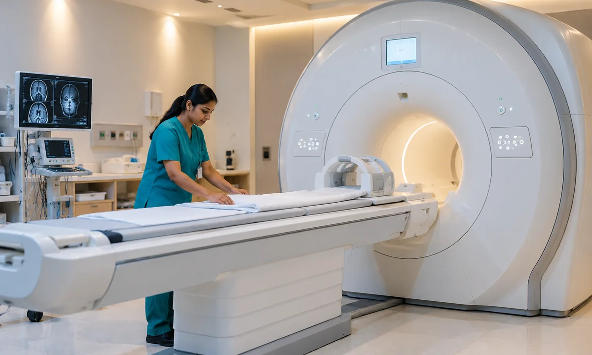
Magnetic resonance imaging (MRI) uses a strong magnetic field and radio waves to produce detailed images. It is often used when clinicians want to assess the brain, spine, joints and other selected soft tissues.
You usually lie on a table that moves into the scanner. The examination can be noisy and you need to remain still. It does not expose you to ionising X-ray radiation. Metal implants and certain devices need careful safety screening; always tell the team about them before entering the scan room.
CT scan: detailed X-ray images, often obtained quickly
Computed tomography (CT) combines multiple X-ray views to create cross-sectional images. It can show many internal structures quickly, which is especially helpful in certain urgent assessments and when a clinician needs detailed views of the chest, abdomen, bones or other body parts.
A CT scanner typically looks like a short, ring-shaped machine. CT does involve ionising radiation, so the referring clinician weighs the expected clinical benefit against potential risks. Some studies use contrast material to show particular structures more clearly.
PREPARING FOR YOUR VISIT
A practical checklist
- Check the exact scan name and body part stated on the referral.
- Tell the imaging team about metal implants, prior contrast reactions or pregnancy where relevant.
- Ask about fasting, a full bladder or other preparation before visiting.
Ultrasound: moving images made with sound waves
Ultrasound (USG) uses high-frequency sound waves to create images of organs and soft tissues. A healthcare professional usually places gel on your skin and moves a handheld probe over the area being examined.
Ultrasound is used for many purposes, including pregnancy monitoring and assessment of the abdomen, pelvis, thyroid and selected blood vessels. Unlike CT, it does not use ionising radiation. Some scans require an empty stomach or a full bladder, depending on what is being examined.
The key differences at a glance
The preparation and appointment duration depend on the exact examination, not only the name of the technology.
- MRI: magnetic field and radio waves; no ionising radiation; important implant-safety checks.
- CT: X-rays; detailed cross-sections, often rapid; a radiation decision is part of test selection.
- Ultrasound: sound waves; no ionising radiation; often uses a gel and handheld probe.
- Contrast: may be needed for selected MRI or CT studies; not every scan needs it.
Questions to ask before your scan
- Which body area is being scanned, and what question is the test meant to answer?
- Will I need fasting, a full bladder or another preparation step?
- Should I mention pregnancy, kidney conditions, allergies or implanted devices?
- Is contrast planned, and do I need to arrange transport?
- When and how should I discuss the report with my treating doctor?
Booking imaging services with Lakhotia Diagnostics
Lakhotia’s published menu includes MRI, CT and ultrasonography, with availability varying by centre. The safest way to plan your visit is to share the exact prescription and preferred location with the booking team before travelling.
If you have sudden severe symptoms or a medical emergency, seek urgent care instead of waiting for a routine scan appointment. Imaging results should always be interpreted with your doctor.
LAKHOTIA DIAGNOSTICS
Need help planning a test?
Confirm test availability, preparation requirements and the most convenient centre before making your appointment.
Frequently asked questions
Is MRI the same as CT?
No. MRI uses magnetic fields and radio waves; CT uses X-rays. They provide different clinical information.
Does ultrasound use radiation?
It uses sound waves, not ionising X-ray radiation.
Will every MRI or CT need contrast?
No. Contrast is used only for selected examinations. Ask the imaging team about your particular order.
How do I know which scan I need?
Your referring clinician should choose the examination based on your symptoms and medical question.
Which scan is best for joint pain or a sports injury?
The choice depends on the suspected problem and examination findings. X-ray, ultrasound, MRI or another test may be appropriate; your clinician decides what is needed.
Sources and further reading
These independent medical resources provide background information for this patient guide. They do not establish which individual investigations are available at each Lakhotia branch.
This guide provides general information, not a diagnosis or a substitute for medical advice. Always follow your clinician's instructions and confirm current test availability and preparation with the centre.